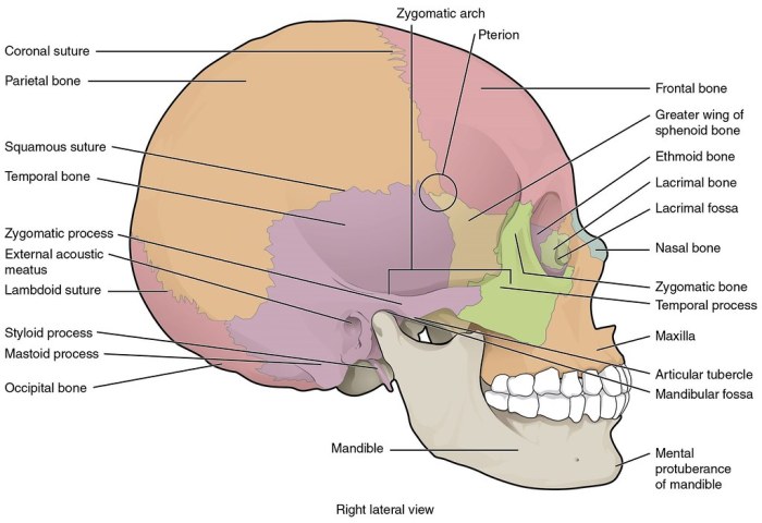
Label the head and neck bones in lateral view provides a comprehensive overview of the skeletal anatomy of the head and neck, offering a detailed examination of the bones, their articulations, and their clinical significance.
This guide explores the anatomical regions of the head and neck, identifying the cranium, facial skeleton, cervical vertebrae, and hyoid bone. It meticulously labels the bones of the cranium, including the frontal, parietal, occipital, temporal, and sphenoid bones, explaining the sutures that connect them.
Anatomical Regions of the Head and Neck

The head and neck are divided into several anatomical regions in lateral view. These regions include the cranium, facial skeleton, cervical vertebrae, and hyoid bone.
Cranium, Label the head and neck bones in lateral view
The cranium forms the bony enclosure of the brain and is composed of eight bones: the frontal, parietal (2), occipital, temporal (2), sphenoid, and ethmoid.
Facial Skeleton
The facial skeleton consists of 14 bones that form the framework of the face. These bones include the nasal, maxillary, zygomatic, lacrimal, mandible, and palatine bones.
Cervical Vertebrae
The cervical vertebrae (C1-C7) form the neck and provide support for the head. Each vertebra has unique features that allow for a wide range of head and neck movements.
Hyoid Bone
The hyoid bone is a small, U-shaped bone located in the midline of the neck. It provides support for the tongue and muscles of the neck.
Helpful Answers: Label The Head And Neck Bones In Lateral View
What are the major anatomical regions of the head and neck?
The major anatomical regions of the head and neck include the cranium, facial skeleton, cervical vertebrae, and hyoid bone.
How many cervical vertebrae are there?
There are seven cervical vertebrae, labeled C1 to C7.
What is the function of the hyoid bone?
The hyoid bone supports the tongue and muscles of the neck.

